Why non-magnetic capacitors matter in medical imaging
By Peter Matthews, Knowles Precision Devices
The quality of an MRI image depends on the homogeneity, or uniformity, of the magnetic field. Component material choice is paramount; even the smallest trace of magnetism inside an MRI scanner can disrupt the field and harm the quality of an MRI image.
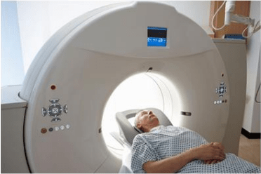
Magnetic resonance imaging (MRI) equipment uses a strong magnetic field and computer-generated radio waves to produce cross-sectional images of soft tissue such as muscle and fat.
These images enable clinicians to investigate and diagnose without the need for more invasive procedures. However, a low-quality image may lead to mistaken diagnoses and, consequently, misguided treatment selections. That said, magnetic resonance applications have very specific needs all the way down to the component level.
MRI basics
When thinking about the working principles behind MRI, it’s important to remember the basics: The MRI machines we are accustomed to are based on the principle of nuclear magnetic resonance (NMR). The name of the phenomenon provides the clue — it has to do with nuclei and magnets.
Magnetic strength is measured in Tesla (T); by extension, Tesla indicates the strength of the MRI’s magnetic field. The 1.5T MRI is one of the more common MRI scanners today, but 3T and 7T machines can produce even higher-resolution images. This level of detail is helpful for diagnosing more unique cases. However, once MRI scanners reach 7T and up, the magnetism is strong enough to cause complications for individuals with implantable devices such as pacemakers.
The molecules that make up the human body contain hydrogen. The nucleus of a hydrogen atom, a single proton, behaves like a magnet with a north and south pole. When a magnetic field is applied, their spins (spin is a property of subatomic particles) arrange uniformly. When a patient is positioned inside the MRI scanner tube, the spins of the protons in their body’s molecules line up, facing the same direction like a marching band practicing on a football field.
When a short, computer-generated RF signal is applied to a portion of the uniform field, those protons receive a “nudge” and break formation. Imagine a scenario in which a stray football is heading for the marching band. After the interruption, the protons (the musicians in our analogy) return to their state of alignment. In the process of realigning, energy is emitted. That energy can be measured and used to distinguish between different types of molecules and their locations. A thorough grasp of MRI requires a deeper dive into quantum mechanics, but beginning to understand the process makes the output that much more amazing.
The catch
An MRI machine is designed to help us identify molecule types and locations based on measuring the behavior of their hydrogen nuclei. However, the quality of an MRI image depends on the homogeneity, or uniformity, of the magnetic field. If there’s any variation, it’s more challenging to detect the impact of an RF signal interruption. Even with the slightest variation, those protons aren’t aligned the same way as the others and won’t respond the same way to stimulus.
These differences confuse the detection algorithms. It would be like some of the musicians in our marching band were already out of step when the football struck. Watching all of this, how would we know where the disturbance caused by the stray ball happened? In practice, excessive signal noise, or random variation in signal intensity, produces granular images. It’s much more challenging for a healthcare professional to rely on them for accurate information.
It’s important for medical device manufacturers to look for high-purity metals that exhibit no measurable magnetism, because magnetic components inside the MRI scanner tunnel can alter the field’s homogeneity. Even the smallest trace of magnetism could affect the quality of the MRI image.
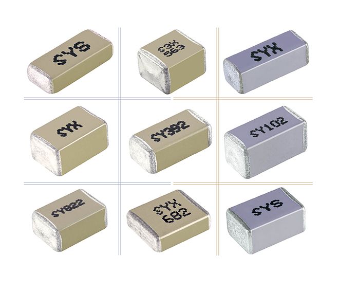
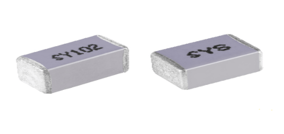
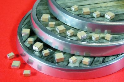
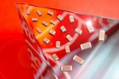
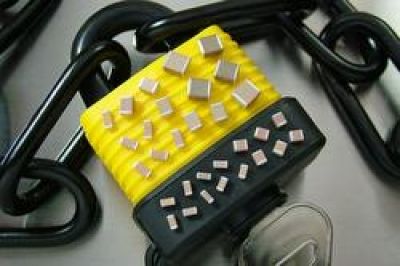
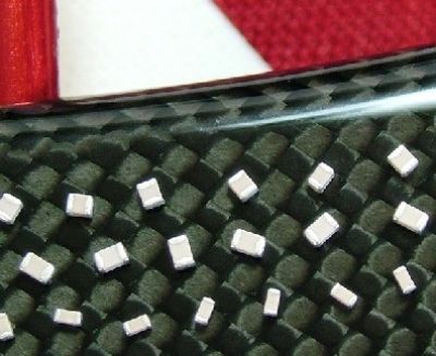
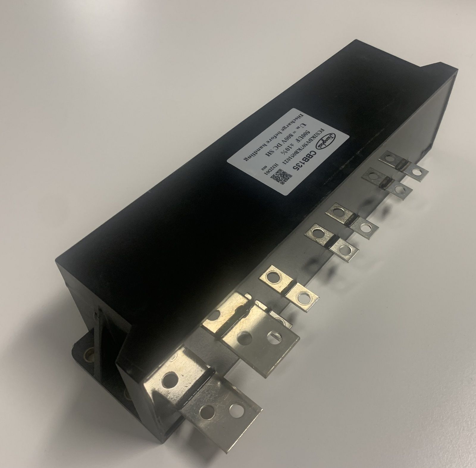
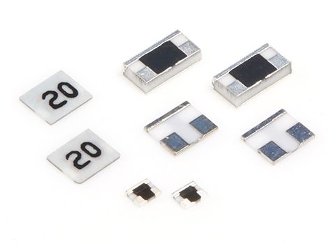
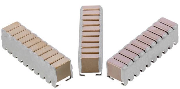
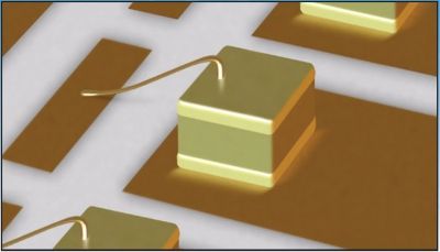
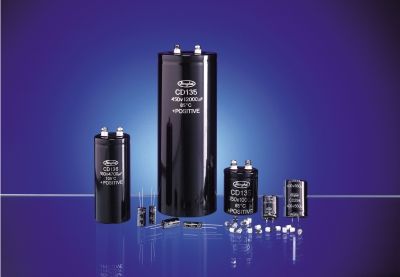
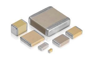
Hardware components including fixed capacitors, trimmer capacitors, inductors, connectors and more must be non-magnetic. Take capacitors, for example: Many capacitors are designed with a nickel barrier finish to maintain solderability. Due to nickel’s magnetic properties, these capacitors are not acceptable for medical applications such as like MRI. Commercial brass, a commonly used material, is also not acceptable for these applications. Coils also require inserts, pins and other special shapes with no measurable magnetism.
This level of care on the component level prevents distortion and minimizes the need for image correction.
Patients, caregivers and healthcare professionals all rely on MRI imaging technology. While components like capacitors are typically viewed as simple or uncomplicated, life-critical applications demand specialized attention in every aspect of design.
Peter Matthews is senior technical marketing manager for Knowles Precision Devices with more than 20 years of experience in technology sales, marketing and product management. Knowles produces highly engineered capacitors and microwave-to-millimeter wave components for use in military, medical, electric vehicle and 5G markets.
Please note: The opinions expressed in this blog post are the author’s only and do not necessarily reflect those of Medical Design and Outsourcing or its employees.

your
local specialist
for